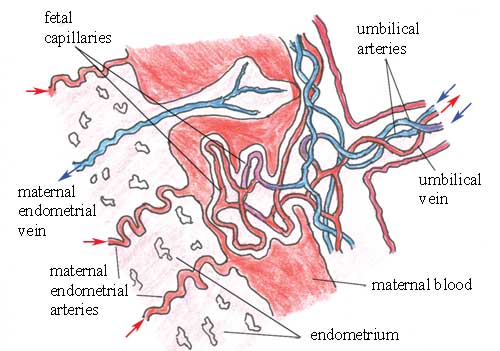
The placental circulation
During fetal development, parts of the placenta project into the maternal endometrium. These small projections are called villi. They produce chemicals that digest and break open the walls of the mother's arteries in the endometrium, so spaces in the placenta fill with oxygenated, nutrient-rich blood from the maternal endometrial arteries.
Villi (singular = villus) is pronounced 'vill-eye'.
However, the pools of maternal blood in the placenta are not in direct contact with the fetal blood. The walls of the smallest fetal arteries and veins in the placenta -- known as capillaries -- remain intact (they are not digested). They are bathed on the outside by maternal blood, which is separated from the fetal blood inside the capillaries by the thickness of the vessel walls.

The fetal capillaries in the placenta are very close to the pool of maternal blood, which has a higher concentration of oxygen and dissolved nutrients than the fetal blood inside the fetal blood vessels.
Think back to your high school biology. What will happen to the oxygen and nutrients in the mother's blood in this situation?
How do you think dissolved waste in the fetal blood is removed as it passes through the placenta?
The placenta has a very large surface area, which facilitates the transport of substances in both directions by diffusion, as described above. Notice that the maternal blood never mixes with the fetal blood -- they are always kept separated by the walls of the fetal blood vessels.